
Abstract
Purpose
Percutaneous nephrolithotomy (PCNL) plays an integral role in treatment of renal stones. Creating percutaneous renal access is the most important and challenging step in the procedure. To facilitate this step, we evaluated our novel mobile augmented reality (AR) system for its feasibility of use for PCNL.
Methods
A tablet computer, such as an iPad\(^{\circledR }\), is positioned above the patient with its camera pointing toward the field of intervention. The images of the tablet camera are registered with the CT image by means of fiducial markers. Structures of interest can be superimposed semi-transparently on the video images. We present a systematic evaluation by means of a phantom study. An urological trainee and two experts conducted 53 punctures on kidney phantoms.
Results
The trainee performed best with the proposed AR system in terms of puncturing time (mean: 99 s), whereas the experts performed best with fluoroscopy (mean: 59 s). iPad assistance lowered radiation exposure by a factor of 3 for the inexperienced physician and by a factor of 1.8 for the experts in comparison with fluoroscopy usage. We achieve a mean visualization accuracy of 2.5 mm.
Conclusions
The proposed tablet computer-based AR system has proven helpful in assisting percutaneous interventions such as PCNL and shows benefits compared to other state-of-the-art assistance systems. A drawback of the system in its current state is the lack of depth information. Despite that, the simple integration into the clinical workflow highlights the potential impact of this approach to such interventions.


Notes
iKlip stand adapter, IK Multimedia Production, http://www.ikmultimedia.com/products/iklip/.
Fig. 2 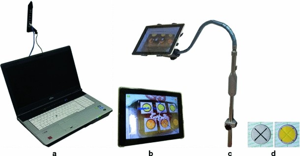
The components of the navigation system: a the navigation server, b Apple iPad as mobile display, c tablet fixation on a flexible endoscope stand, d radio-dense navigation markers, optionally attached with color labels for a more robust localization
Braun Melsungen AG, http://www.bbraun.de/.
Rebeck patient markers, Fobeck GbR, http://fobeck.com/cms2/en.
For a detailed specification, please refer to the manufacturer‘s website http://www.siemens.com.

References
Romero V, Akpinar H, Assimos DG (2010) Kidney stones: a global picture of prevalence, incidence, and associated risk factors. Rev Urol 12(2–3):86–96
Skolarikos A, Alivizatos G, de la Rosette JJ (2005) Percutaneous nephrolithotomy and its legacy. Eur Urol 47(1):22–28
Michel MS, Trojan L, Rassweiler JJ (2007) Complications in percutaneous nephrolithotomy. Eur Urol 51(4):899–906
Andonian S, Scoffone C, Louie MK, Gross AJ, Grabe M, Daels F, Shah HN, De La Rosette J (2012) Does imaging modality used for percutaneous renal access make a difference? a matched case analysis. J Endourol 27(1):24–28
Nicolau S, Soler L, Mutter D, Marescaux J (2011) Augmented reality in laparoscopic surgical oncology. Surg Oncol 20(3): 189–201
Maier-Hein L, Tekbas A, Seitel A, Pianka F, Müller SA, Satzl S, Schawo S, Radeleff B, Tetzlaff R, Franz AM, Müller-Stich BP, Wolf I, Kauczor H-U, Schmied BM, Meinzer H-P (2008) In vivo accuracy assessment of a needle-based navigation system for CT-guided radiofrequency ablation of the liver. Med Phys 35(12):5385–5396
Fichtinger G, Deguet A, Fischer G, Iordachita I, Balogh E, Masamune K, Taylor RH, Fayad LM, de Oliveira M, Zinreich SJ (2005) Image overlay for CT-guided needle insertions. Comput Aided Surg 10(4):241–255
Song DY, Burdette EC, Fiene J, Armour E, Kronreif G, Deguet A, Zhang Z, Iordachita I, Fichtinger G, Kazanzides P (2011) Robotic needle guide for prostate brachytherapy: clinical testing of feasibility and performance. Brachytherapy 10(1):57–63
Wein W, Brunke S, Khamene A, Callstrom MR, Navab N (2008) Automatic CT-ultrasound registration for diagnostic imaging and image-guided intervention. Med Image Anal 12(5):577–585
Wood BJ, Kruecker J, Abi-Jaoudeh N, Locklin JK, Levy E, Xu S, Solbiati L, Kapoor A, Amalou H, Venkatesan AM (2010) Navigation systems for ablation. J Vasc Interv Radiol 21(8 Suppl):257–263
Lazarus J, Williams J (2011) The locator: novel percutaneous nephrolithotomy apparatus to aid collecting system puncture-a preliminary report. J Endourol 25(5):747–750
Huber J, Wegner I, Meinzer HP, Hallscheidt P, Hadaschik B, Pahernik S, Hohenfellner M (2011) Navigated renal access using electromagnetic tracking: an initial experience. Surg Endosc 25(4):1307–1312
Ritter M, Rassweiler MC, Hacker A, Michel MS (2012) Laser-guided percutaneous kidney access with the Uro Dyna-CT: first experience of three-dimensional puncture planning with an ex vivo model. World J Urol. 30:1–5
Baumhauer M, Simpfendörfer T, Stich BM, Teber D, Gutt C, Rassweiler J, Meinzer H-P, Wolf I (2008) Soft tissue navigation for laparoscopic partial nephrectomy. Int J Comput Assist Radiol Surg 3:307–314
Maier-Hein L, Franz AM, Fangerau M, Schmidt M, Seitel A, Mersmann S, Kilgus T, Groch A, Yung K, dos Santos TR, Meinzer H-P (2011) Towards mobile augmented reality for on-patient visualization of medical images. In: Handels H, Ehrhardt J, Deserno TM, Meinzer H-P, Tolxdorff T (eds) Bildverarbeitung für die Medizin. Springer, Berlin, pp 389–393
Rassweiler JJ, Müller M, Fangerau M, Klein J, Goezen AS, Pereira P, Meinzer HP, Teber D (2012) iPad-assisted percutaneous access to the kidney using marker-based navigation: initial clinical experience. Eur Urol 61(3):628–631
Wolf I, Vetter M, Wegner I, Böttger T, Nolden M, Schöbinger M, Hastenteufel M, Kunert T, Meinzer H-P (2005) The medical imaging interaction toolkit. Med Image Anal 9(6):594–604
Fangerau M (2011) Medical imaging interaction toolkit for mobile devices. Master’s thesis, Hochschule Mannheim, University of Applied Sciences
Müller M, Groch A, Baumhauer M, Maier-Hein L, Teber D, Rassweiler J, Meinzer H-P, Wegner I (2012) Robust and efficient fiducial tracking for augmented reality in HD-laparoscopic video streams. In: DRH III, Wong KH, (eds) SPIE medical imaging 2012: visualization, image-guided procedures, and modeling, vol 8316, No 1. SPIE, p 83161M
Zhang Z (2000) A flexible new technique for camera calibration. IEEE T Pattern Anal 22:1330–1334
Lowe DG (1991) Fitting parameterized three-dimensional models to images. IEEE Trans Pattern Anal Mach Intell 13:441–450
DeMenthon DF, Davis LS (1995) Model-based object pose in 25 lines of code. Int J Comput Vis 15:123–141
Lu C-P, Hager GD, Mjolsness E (2000) Fast and globally convergent pose estimation from video images. IEEE Trans Pattern Anal Mach Intell 22:610–622
Lepetit V, Moreno-Noguer F, Fua P (2008) EP\(n\)P: an accurate o(\(n\)) solution to the P\(n\)P problem. Int J Comput Vis 81(2):155–166
Sarkis M, Diepold K (2012) Camera-pose estimation via projective Newton optimization on the manifold. IEEE Trans Image Process 21(4):1729–1741
Li S, Xu C, Xie M (2012) A robust o(n) solution to the perspective-n-point problem. IEEE Trans Pattern Anal Mach Intell. 34(7):1444–1450
Fischler MA, Bolles RC (1981) Random sample consensus: a paradigm for model fitting with applications to image analysis and automated cartography. Commun ACM 24(6):381–395
Nocedal J, Wright SJ (2006) Numerical optimization, 2nd edn. ser. Springer series in operations research and financial engineering. Springer, New York
Holloway RL (1997) Registration error analysis for augmented reality. Presence Teleoper Virtual Environ 6(4):413–432
Baumhauer M (2008) Real-time compensation of organ motion for augmented reality in laparoscopic surgery. Ph.D. dissertation, Ruprecht-Karls University Heidelberg
Simpfendörfer T, Baumhauer M, Müller M, Gutt CN, Meinzer HP, Rassweiler JJ, Guven S, Teber D (2011) Augmented reality visualization during laparoscopic radical prostatectomy. J Endourol 25:1841–1845
Laganière R (2011) OpenCV 2 computer vision application programming cookbook. Packt Publishing, Birmingham
Miller NL, Lingeman JE (2007) Management of kidney stones. BMJ 334(7591):468–472
Ritter M, Siegel F, Krombach P, Martinschek A, Weiss C, Hacker A, Pelzer AE (2012) Influence of surgeon’s experience on fluoroscopy time during endourological interventions. World J Urol 31(1): 183–187
Seitel A, Maier-Hein L, Schawo S, Radeleff B, Müller SA, Pianka F, Schmied BM, Wolf I, Meinzer H-P (2007) In-vitro evaluation of different visualization approaches for computer assisted targeting in soft tissue. In: Lemke H, Inamura K, Doi K, Vannier M, Farman A (eds) International journal of Computer Assisted Radiology and Surgery. Berlin (Germany), pp 188–190, June 2007
Karami H, Rezaei A, Mohammadhosseini M, Javanmard B, Mazloomfard M, Lotfi B (2010) Ultrasonography-guided percutaneous nephrolithotomy in the flank position versus fluoroscopy-guided percutaneous nephrolithotomy in the prone position: a comparative study. J Endourol 24(8):1357–1361
Agarwal M, Agrawal MS, Jaiswal A, Kumar D, Yadav H, Lavania P (2011) Safety and efficacy of ultrasonography as an adjunct to fluoroscopy for renal access in percutaneous nephrolithotomy (PCNL). BJU Int 108(8):1346–1349
Maier-Hein L, Walsh CJ, Seitel A, Hanumara NC, Shepard J-A, Franz AM, Pianka F, Müller SA, Schmied B, Slocum AH, Gupta R, Meinzer H-P (2009) Human vs. robot operator error in a needle-based navigation system for percutaneous liver interventions. In: SPIE medical imaging 2009: visualization, image-guided procedures, and modelling, vol 7261, p 72610Y (12 p)
Franz A, März K, Hummel J, Birkfellner W, Bendl R, Delorme S, Schlemmer H-P, Meinzer H-P, Maier-Hein L (2012) Electromagnetic tracking for us-guided interventions: standardized assessment of a new compact field generator. Int J Comp Assist Radiol Surg 7:1–6
Yaniva Z, Wilson E, Lindisch D, Cleary K (2009) Electromagnetic tracking in the clinical environment. Med Phys 36(3):876–892
Lee T, Hollerer T (2009) Multithreaded hybrid feature tracking for markerless augmented reality. IEEE Trans Vis Comput Graph 15(3):355–368
Grimm R, Bauer S, Sukkau J, Hornegger J, Greiner G (2012) Markerless estimation of patient orientation, posture and pose using range and pressure imaging. Int J Comput Assist Radiol Surg 1:1–9
Newcombe RA, Davison AJ, Izadi S, Kohli P, Hilliges O, Shotton J, Molyneaux D, Hodges S, Kim D, Fitzgibbon A (2011) Kinectfusion: real-time dense surface mapping and tracking. In: Mixed and augmented reality (ISMAR), 2011 10th IEEE international symposium on, pp. 127–136, Oct 2011
Mersmann S, Gergel I, Seitel A, Gaa J, Wegner I, Meinzer H-P, Maier-Hein L (2011) Microsoft kinect controller as intra-operative imaging modality. Int J Comput Assist Radiol Surg 6(Suppl 1): 251–252
Seitel A, Engel M, Sommer CM, Radeleff BA, Essert-Villard C, Baegert C, Fangerau M, Fritzsche KH, Yung K, Meinzer H-P, Maier-Hein L (2011) Computer-assisted trajectory planning for percutaneous needle insertions. Med Phys 38(6):3246–3259
Acknowledgments
The presented work was conducted within the setting of the “Research group 1126: Intelligent Surgery-Development of new computer-based methods for the future workplace in surgery” funded by the German Research Foundation (DFG). Furthermore, we want to thank the staff in the urological departments of our partner hospitals for the support during our experiments. The presented software was developed as part of the Medical Imaging Interaction Toolkit (MITK, http://www.mitk.org).
Conflict of Interest
None.
Author information
Authors and Affiliations
Corresponding author
Electronic supplementary material
Below is the link to the electronic supplementary material.
Rights and permissions
About this article
Cite this article
Müller, M., Rassweiler, MC., Klein, J. et al. Mobile augmented reality for computer-assisted percutaneous nephrolithotomy. Int J CARS 8, 663–675 (2013). https://doi.org/10.1007/s11548-013-0828-4
Received:
Accepted:
Published:
Issue Date:
DOI: https://doi.org/10.1007/s11548-013-0828-4